'%3e%3cg%20transform='translate(76.124%2037.922)'%20style='isolation:isolate'%3e%3cpath%20d='M21.985-77.7l-5.958,8.856L10.07-77.7H7.77v16.1h2.3V-73.445l5.543,8.166h.828l5.543-8.166V-61.6h2.3V-77.7Zm9.408,4.163a7.286,7.286,0,0,0-4.9,1.817l1.472,1.127a5.462,5.462,0,0,1,3.013-1.035c2.415,0,2.783,1.38,2.783,3.151H31.876c-1.633,0-6.073.092-6.073,3.749,0,2.093,1.932,3.427,4.14,3.427a4.564,4.564,0,0,0,3.818-1.909v1.61h2.093v-7.039C35.855-71.42,34.751-73.537,31.393-73.537Zm2.369,8.6A3.655,3.655,0,0,1,30.4-63c-1.863,0-2.438-.92-2.438-1.817,0-2.047,2.829-2.024,4.991-2.024h.805Zm9.891-8.6a4.157,4.157,0,0,0-3.611,1.909v-1.633H37.949V-61.6h2.093v-6.694a3.131,3.131,0,0,1,3.105-3.266c2.185,0,2.875,1.633,2.875,3.266V-61.6h2.093V-68.89A4.521,4.521,0,0,0,43.653-73.537Zm11.869,2.277v-2H52.831V-75.7H50.738v2.438H48.921v2h1.817v5.866c0,2.553,1.127,3.91,3.657,3.91a5,5,0,0,0,1.127-.115v-1.84c-.253.023-.506.046-.851.046-1.817,0-1.84-1.1-1.84-2.76v-5.106Zm6.648-2.277a7.286,7.286,0,0,0-4.9,1.817l1.472,1.127a5.462,5.462,0,0,1,3.013-1.035c2.415,0,2.783,1.38,2.783,3.151H62.653c-1.633,0-6.073.092-6.073,3.749,0,2.093,1.932,3.427,4.14,3.427a4.564,4.564,0,0,0,3.818-1.909v1.61h2.093v-7.039C66.632-71.42,65.528-73.537,62.17-73.537Zm2.369,8.6A3.655,3.655,0,0,1,61.181-63c-1.863,0-2.438-.92-2.438-1.817,0-2.047,2.829-2.024,4.991-2.024h.805ZM68.725-77.7v16.1h2.093V-77.7Zm12.237,4.439-3.2,8.419-3.588-8.419H71.854L76.73-62.2l-2.07,5.037h2.254l6.372-16.1Zm7.361,4.9c-1.334-.276-2.944-.368-2.944-1.679,0-.966.851-1.61,2-1.61a2.137,2.137,0,0,1,2.208,1.334l1.955-.529a3.832,3.832,0,0,0-3.979-2.691c-2.53,0-4.3,1.518-4.3,3.657,0,1.725.943,2.783,3.128,3.266,1.61.368,3.565.368,3.565,1.725,0,1.035-.851,1.656-2.369,1.656-1.886,0-2.507-.828-2.691-1.426l-2.047.69c.46,1.748,2.392,2.668,4.692,2.668,3.105,0,4.577-1.725,4.577-3.8C92.118-66.843,91.175-67.763,88.323-68.361Z'%20transform='translate(-7.77%2077.7)'%20fill='%23efefef'/%3e%3c/g%3e%3cg%20transform='translate(37.428%2032.714)'%3e%3cg%20transform='translate(0%200)'%20clip-path='url(%23a)'%3e%3cpath%20d='M27.251,14.507,22.633,3.938A7.662,7.662,0,0,0,15.938,0H0L6.428,6.428a2.865,2.865,0,0,0,2.026.839h8.092A7.443,7.443,0,0,1,21.56,9.209Z'%20transform='translate(0%200)'%20fill='%23fcc45c'/%3e%3cpath%20d='M99.681,168.129l-5.915-5.5a5.363,5.363,0,0,0-5.4-.659l-10.231,4.449,5.921,2.332a2.005,2.005,0,0,0,1.535-.027l5.195-2.259a5.21,5.21,0,0,1,3.761-.153Z'%20transform='translate(-73.605%20-152.158)'%20fill='%23fcc45c'/%3e%3cpath%20d='M202.96,271.253l-5.331-1.877a3.754,3.754,0,0,0-3.649,1.084l-5.326,5.711,4.452-.155a1.4,1.4,0,0,0,.978-.446l2.7-2.9a3.647,3.647,0,0,1,2.371-1.148Z'%20transform='translate(-177.709%20-253.644)'%20fill='%23fcc45c'/%3e%3cpath%20d='M311.272,336.306l-3.946.283a2.628,2.628,0,0,0-2.04,1.714l-1.825,5.153,2.815-1.342a.982.982,0,0,0,.5-.559l.927-2.616a2.553,2.553,0,0,1,1.2-1.4Z'%20transform='translate(-285.855%20-316.795)'%20fill='%23fcc45c'/%3e%3c/g%3e%3c/g%3e%3c/g%3e%3c/svg%3e)
Light, fast, high-performance AI
solutions for scientific and
industrial image analysis
Mantalys
Visionary Intelligence
Mantalys creates light, fast, high-performance AI solutions for image analysis by unlocking the power of both Evolutionary Computation and Deep Learning approaches.
We offer custom algorithm design services and intuitive software packages for scientific imaging.
MantaPlex
Multiplex Spatial Image Analysis
An intuitive software platform that leverages artificial intelligence for high-dimensional spatial analysis of multiplexed fluorescent images
MantaPlex offers:
- High-dimensional spatial analysis for multiplexed whole slide images
- Fast, local image processing (no cloud-based computing or data storage)
- State-of-the-art algorithms for cell segmentation, classification, and spatial analysis
- Intuitive user interface and expert-designed guided workflow
- An interactive Results Dashboard with easy export of summary or single-cell data
A guided workflow you can trust: designed for biomedical researchers without informatics experience, MantaPlex offers users both the freedom to explore different analysis approaches and the step-by-step guidance to produce reproducible results with confidence.
Our software is equipped with rigorously validated pre-trained algorithms for common image analysis tasks. Users can experiment with tunable analysis parameters and view results immediately using the Results Dashboard.
Interested in exclusive pre-release access to MantaPlex?
Sign up as an Early Innovator to benefit from early access to our software, supported by one-on-one training sessions and direct engagement with the scientists and developers at Mantalys.
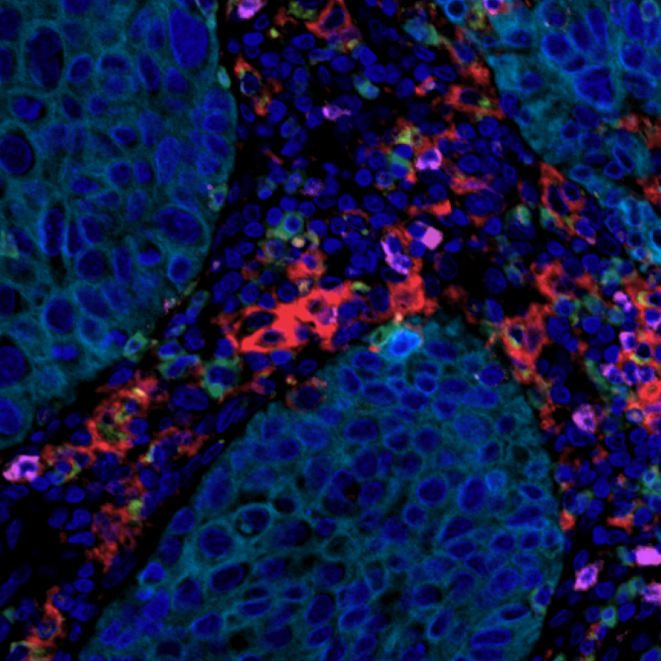
Tissue visualization and annotation
Intuitive user-friendly tools for visualizing and annotating highly multiplexed tissue from whole-slide images
With our intuitive tissue visualization and annotation tools, you can:
- Import whole-slide images and select tissue regions of interest
- Adjust channels, remove background, and use custom colours for your high-plex markers using our channel manager
- Use our customizable grid view for single-channel inspection and visual validation prior to downstream data analysis
- Annotate tissue structures and regions of interest to perform compartment-specific downstream analyses
- Facilitate navigation of large tissue sections by using drag-and-drop pins to landmark important features
- Create customized multi-layer images and export snapshots of annotated tissue with just a few clicks
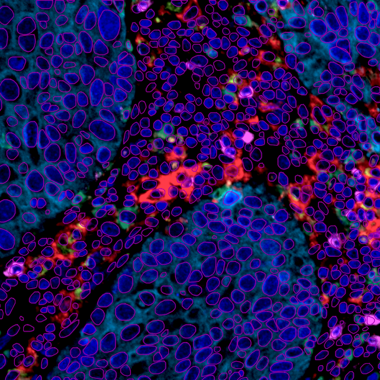
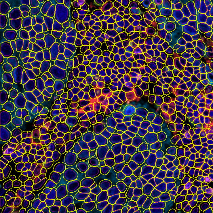
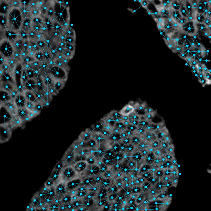
High-dimensional single-cell analysis
Use state-of-the-art algorithms to automatically segment and classify cell types and phenotypes
With MantaPlex you can:
- Detect nuclei and individual cells with high precision using pre-trained state-of-the-art segmentation algorithms
- Identify cell types automatically using rule-based cell classification informed by user-generated cell classification rules and manually-tuned positivity thresholds
- Generate exportable single-cell feature tables to assess biomarker expression at the single-cell level
- Assess and compare cellular composition across previously identified tissue compartments
- Generate U-MAPs based upon single-cell feature extraction to visualize high-dimensional datasets
- Create customized multi-layer images and export snapshots of segmented and classified cells
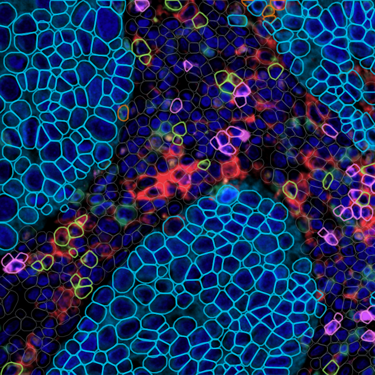
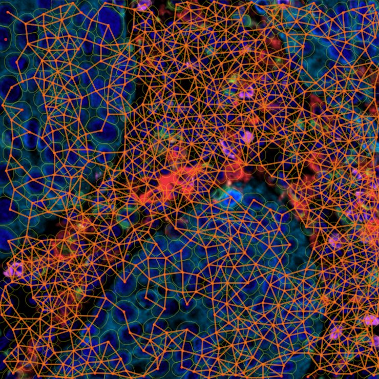
Multiplex spatial image analysis
Use state-of-the-art algorithms to identify spatial relationships between cells
Examine spatial relationships between different cell populations within the complex tissue microenvironment:
- Understand relationships between cell types by performing proximity profile and nearest neighbour analyses
- Export biomarker expression data stratified according to spatial context
- Inspect cell:cell interactions using our intuitive visualization tools
- Create customized multi-layer images and export snapshots of spatial analyses using just a few clicks
Services
How we can help you
We collaborate with domain experts to develop customized image analysis solutions to address their most challenging image processing and analysis needs.
We offer:
- One-on-one consultations with image analysis experts
- State-of-the-art Deep Learning and Evolutionary Computation approaches, as well as a library of custom algorithms designed in-house
- Flexible delivery to integrate solutions into different workflows (e.g. compiled code, deployable models)
Get in touch
Contact us here to request more information on how we can solve your image analysis challenges.
Partnership Inquiries
By becoming a partner, you will gain access to our extensive network, innovative resources, and dedicated support team. Together, we can achieve greater success and make a significant impact in our industry.